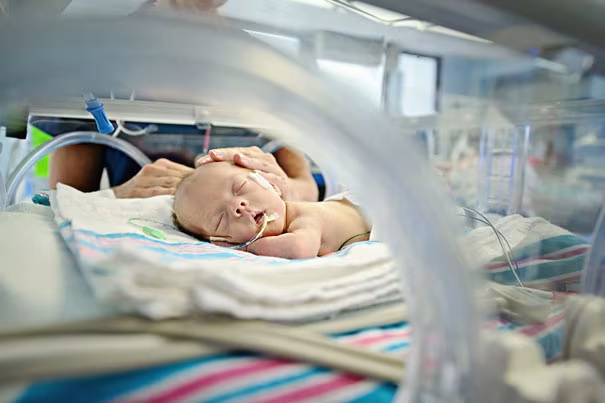
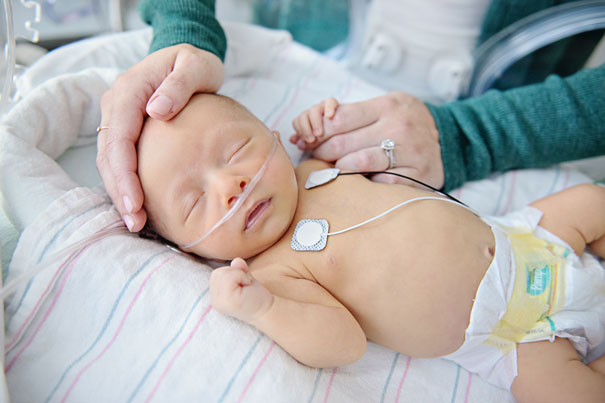
Neonatal Intensive Care Unit (NICU): Common Problems
Here's an alphabetized neonatal diagnosis list of the most common conditions and treatments that may be part of your infant's early life in the NICU and beyond. Whenever you hear your baby’s doctors and nurses mention a new term, ask for an explanation, or at least write it down so you can consult this list later.
For more information to help familiarize yourself with the NICU, be sure to check out the related guides that cover staff, equipment, and tests.
Common Diagnoses in the NICU
Below is a list of common neonatal disorders you might encounter during your baby's stay in the NICU.
ABO incompatibility. A condition that arises when a mother with type-O blood has an infant with type-A or type-B blood. Her body makes proteins that cross the placenta and cause a rapid breakdown of the blood in a fetus or newborn. This creates jaundice and anemia in the infant. ABO incompatibility is less severe than Rh incompatibility (see Rh incompatibility below).
Anemia. An insufficient amount of red blood cells, often found in premature babies. Normally, the fetus stores iron during the later months of pregnancy and uses it after birth to make red blood cells, but infants born early may not have had enough time to store iron. Loss of blood from frequent blood tests, as well as a delay in bone marrow activation, can also cause anemia. Anemic NICU babies may be treated with dietary iron supplements, drugs that increase red blood cell production, or, in some cases, a blood transfusion.
Apnea. A stoppage in breathing. NICU cases often involve premature babies who sometimes do not breathe regularly. A baby may take a long breath, then a short one, and then pause for 5 to 10 seconds before starting to breathe normally. This is called periodic breathing. It usually is not harmful, and the baby will outgrow it. However, if a pause lasts longer than 15 seconds, it usually requires some intervention and treatment. Medication may be used to regulate the baby's breathing.
Asphyxia. The poor exchange of blood gases, with low oxygen and high carbon dioxide. This can occur before or after birth. Infants can tolerate this better than older children or adults, but severe asphyxia can cause problems in several organs. Often, the term is used to mean hypoxia or anoxia, low or absent oxygen.
Aspiration. Breathing into the lungs of some substance that shouldn't be there, such as amniotic fluid, meconium, or formula. This may cause aspiration pneumonia.
Bradycardia. Slow heart rate. This can indicate a problem such as infection or seizures, or it can follow or accompany apnea. If frequent, additional testing and treatment may be needed. When occurring with apnea, the condition is sometimes referred to as "A's and B's. " It can sometimes be resolved with just a little stimulation of the infant.
Breathing problems. Breathing problems are common in NICU cases, often seen in premature babies when their lungs are not fully developed and the brain systems that regulate their breathing may still be immature. Full-term babies also can develop breathing problems due to complications of labor and delivery, physical defects, and infections. These problems may be helped by medicines, the use of a respirator or other NICU monitoring devices to support or assist the baby's breathing, or a combination of interventions.
Bronchopulmonary dysplasia (BPD). BPD is a common NICU diagnosis that occurs when there have been serious breathing problems after birth, and the lungs and bronchial tubes have sustained some damage and scarring. Babies with severe respiratory distress syndrome (see Respiratory distress syndrome below) who require prolonged treatment with mechanical ventilation and oxygen are at risk for BPD. Usually, the lungs heal during the first years of life, though sometimes BPD can persist and become an asthma-like condition. BPD may require special treatment during hospitalization and continuing at home until the lungs heal adequately. Fluids need to be limited for infants with BPD, making nutrition a big challenge. Pneumonia is common when there is a chronic lung condition such as BPD.
Cerebral palsy. A condition involving disturbances in motor tone and the smooth control and coordination of motor movements. It results from permanent but non-progressive damage to motor areas of the brain before or around the time of birth. Preemies are more likely than full-term infants to develop CP. Because many preemies have motor-system abnormalities that disappear over time, cerebral palsy may go undiagnosed until a year or more after birth. The range of abnormalities goes from subtle to severe. Intellectual functioning may be normal.
Cerebrospinal fluid (CSF). The fluid around the brain and spinal cord. Taking a sample of this for testing is called a lumbar puncture, or spinal tap.
Chest physiotherapy (CPT or Chest PT). Tapping, moving, and vibrating the chest to loosen secretions so they can be cleared out of the lungs, often by suctioning with a catheter.
Coarctation of the aorta. A narrowing of part of the aorta, the large artery that connects the heart to the rest of the body, so that blood doesn't flow evenly. A surgeon may need to repair this. Sometimes, the narrowed area can be widened by inflating a balloon on the tip of a catheter inserted through an artery.
Cyanosis. Bluish discoloration of the lips, gums, or extremities. This may develop if the oxygen content of an infant's blood is too low because of heart, lung, or brain problems. Blood oxygen tests can determine how serious the problem is, while other tests can help to define the cause. (Note: All young infants will display bluish fingers and toes—without central cyanosis or discoloration of the lips—when they are cold. This type of cyanosis is generally of no concern.)
Desaturation. A reduction of the level of oxygen in the blood, often used as a verb, as in "He's desatting. " More oxygen or more respiratory support is usually needed to correct the situation.
Feeding difficulties. Swallowing incoordination, fatigue, or poor gag or sucking reflexes, which may necessitate special methods of feeding until the baby is ready for the breast or bottle. Babies who are very small or sick are initially fed intravenously (through a vein). A tiny, plastic tube or catheter can be placed in a vein in the baby's hand, foot, scalp, or belly button to deliver sugar (glucose) and essential nutrients for several days. A longer-lasting tube called a central venous catheter can be placed into a neck or arm vein and threaded into a deep vein in the chest. It can remain in place for several weeks to provide nutritional support and deliver medication.
As soon as the baby is strong enough, they will be fed breast milk or formula through a tube placed through the nose or mouth into the stomach (a nasogastric tube) or into the intestines (a nasojejunal tube). This is called gavage feeding. In gavage feeding, the tube may either be left in place or inserted at each feeding. Inserting a tube should not bother the baby too much because babies this small generally do not gag. Sometimes the stomach and intestines don't move food along as they should, so some is left over, and this is called residuals. When the baby can suck and swallow effectively, breast milk or formula feedings are slowly introduced, and the gavage feedings are eventually stopped.
Gastrostomy tube/button (G-tube). A tube or opening directly into the stomach from the abdominal wall. This allows for prolonged feeding of infants who cannot feed orally. Special formulas may be given continuously by pump infusion or dripped in periodically at mealtimes.
Heart failure. The inability of the heart to maintain adequate pumping, either because of a problem with the heart itself or with the lungs or kidneys. A salt imbalance can cause heart failure as well. Typically, the heart will look enlarged on an X-ray. One common drug given to stimulate the heart is digitalis, but many other medicines and treatments are also used in NICU hospitals.
Heart murmur. An extra heart sound caused by rapid or wayward blood flow through the heart. It may indicate a heart problem, anemia, or infection. Many infants have "innocent murmurs," which are not cause for concern because blood flows more rapidly through the infant heart. Many preemies will have a small heart murmur up until their first birthday or beyond with no need for any treatment.
Heart valve abnormalities. The narrowing, closing, or blockage of a heart valve, which prevents blood from flowing smoothly. Some babies may require placement of a shunt (artificial graft) to allow blood to bypass the blockage until the baby is big enough to have the valve repaired or replaced.

Hernia/hydrocele. Two bulges in the groin area. When the lower abdominal wall isn't completely sealed off, a membrane sac from the abdomen remains in the scrotum, creating an inguinal hernia, which often requires a simple surgical repair. A second reason for a bulge is fluid around the testes, called a hydrocele. It may look like a hernia but virtually never requires any treatment except time.
Hypoglycemia. Low blood sugar. Hypoglycemia is a condition frequently seen in NICU cases, occurring right after birth if there is a delay in the infant's ability to mobilize stored glucose. Small infants may have reduced body stores to begin with. Stressed infants may use glucose more quickly than they can replace it. Babies born to mothers with diabetes are at greater risk for hypoglycemia because they have an excess of circulating insulin. A very small sample of blood from the baby's heel can check glucose levels quickly. Early feeding and intravenous glucose help to prevent and treat hypoglycemia.
Hypotension, hypertension. Abnormal blood pressure (too low or too high). This may be due to any of several concerns that require different interventions. Your baby's blood pressure will be monitored nearly continuously if they are very sick, with the reading shown on a monitor at their bedside. Medicine, adjustment of intravenous fluids, or other treatments may be needed.
Hypothermia. Low body temperature. Babies who are born too small and too soon often have trouble controlling their body temperature. Unlike healthy, full-term babies, they do not have enough body fat to prevent the loss of heat. Infants in the NICU are placed in a baby incubator or warmer right after birth to help control their temperature. A tiny sensor taped to the baby's stomach will measure their temperature and regulate the heat in the incubator or through the radiant heater. Very small babies will be covered by a cellophane blanket, helping them maintain their temperature and avoid water loss through very thin skin. A hat helps reduce heat loss from the blood-vessel-rich scalp. A baby will grow faster if they maintain a normal body temperature. In fact, one criterion for release from the hospital is the ability to maintain temperature.
Intrauterine growth retardation (IUGR). A condition in which a baby grows more slowly than usual in utero and is smaller than normal for their gestational age at birth (termed SGA, or small for gestational age). There are multiple causes, and testing may be needed to evaluate the condition.
Intraventricular hemorrhage (IVH). Bleeding into the cavities (ventricles) of the brain, a condition most common in the smallest premature babies. An ultrasound examination can show whether a baby has had brain bleeding and how severe it is, noted by a grade from 1 to 4. Most brain bleeds are mild (grades 1 and 2) and resolve themselves with no or few lasting problems. More severe bleeds can cause difficulties for the baby during hospitalization and possible problems in the future. Large bleeds may block the normal outflow of brain fluid, creating a backup of pressure and expansion of the ventricles. This is called hydrocephalus.
Jaundice (hyperbilirubinemia). An excess of bilirubin, which comes from the breakdown of red blood cells. Jaundice is common in premature babies and in infants who have blood-type incompatibilities with their mothers. Babies with jaundice have a yellowish tinge to their skin and eyes, but the condition is worrisome only when the liver is too immature or sick to clear the bilirubin from the blood. If the bilirubin level gets too high, it can cause serious brain problems. For this reason, a baby's bilirubin level is checked frequently. If it gets too high, they will be treated with special blue lights ("bili lights") that help the body break down and eliminate bilirubin. During this treatment, called phototherapy, the baby wears a minimum of clothing as well as blinders to protect their eyes. Occasionally, a baby will need a special type of blood transfusion called an exchange transfusion to reduce very high bilirubin levels. In this procedure, some of the baby's blood is removed and replaced with blood from a donor.
Laryngomalacia/tracheomalacia. A softening of the vocal cords (larynx) or the windpipe (trachea), making them floppy enough to interfere with breathing.
Meconium aspiration. Breathing in the infant's first bowel movement just prior to or during delivery. This sticky, green-black, tar-like material is passed if the infant is overly stressed in labor or at delivery. When it gets into the lungs, meconium produces a kind of pneumonia that prevents good gas exchange.
Necrotizing enterocolitis (NEC). NEC is a serious NICU diagnosis that occurs when the bowel becomes damaged due to decreased blood supply or poor oxygenation. Bacteria normally present in the bowel invade the damaged area, causing more damage. A baby with NEC develops feeding problems, abdominal swelling, and other complications, even rupture of the bowel. They will be fed intravenously while their bowel heals. Sometimes, damaged sections of the intestine must be surgically removed.
Patent ductus arteriosus (PDA). The most common heart problem in premature babies. Before birth, much of the fetus's blood bypasses the lungs through a passageway called the ductus arteriosus. This passageway normally closes soon after birth. If it doesn't close, too little blood flows into the lungs, making the heart work harder. In some cases, drug treatment can help close the passageway. If that doesn't work, surgery may be necessary.
Periodic breathing. Irregular breathing in which pauses of 10 to 20 seconds may occur normally in young infants, particularly preemies. It may be linked to a slowing of the heart or a drop in blood oxygen level. If pauses in breathing are longer than 15 seconds, the condition is considered abnormal and is then known as apnea.
Periventricular leukomalacia (PVL). Damage to areas of the brain around the ventricles, including a possible loss of tissue with cyst formation. This is usually seen with the higher grades or more serious cases of intraventricular hemorrhage (bleeding). It can also develop without bleeding in very small or very ill infants. An ultrasound, CT scan, or MRI will show this condition. If persistent, cerebral palsy may result.
Persistent pulmonary hypertension of the newborn (PPHN). Elevated blood pressure in the lungs, which prevents babies from breathing properly. At birth, the blood vessels in the lungs normally relax and allow blood to flow through them. In babies with PPHN, this response does not occur, which leads to a lack of oxygen in the blood. Babies with PPHN often have other problems (such as heart defects) or have suffered from birth complications. They often need a ventilator (respirator) to help them breathe. A treatment with nitric oxide (a gas administered through a tube in the windpipe) may help the blood vessels in the lungs to relax and may improve breathing.
Pneumonia. A lung infection that is common in premature and sick newborns. Doctors may suspect pneumonia if the baby has difficulty breathing, shows changes in their rate of breathing, or has an increased number of apnea episodes. An X-ray will show the area of pneumonia. Antibiotics are usually given after lung secretions are obtained to find out which germ is the culprit.
Pneumothorax. Air in the chest cavity. This occurs when a very stiff lung (due to respiratory distress syndrome, pneumonia, or other serious lung condition) blows out a bleb, or bubble, much like the blowout of a tire, and air leaks into the chest cavity. Stiff lungs and the need for high pressures on a ventilator may occur with RDS (see Respiratory distress syndrome below), meconium aspiration, or any kind of pneumonia. A pneumothorax sometimes requires the placement of a tube into the chest to suck out the air until the leak repairs itself.
Polycythemia. A condition in which there are too many red blood cells, which makes the blood thick, or hyperviscous. The opposite of anemia, polycythemia may require an exchange transfusion.
Reflux. The return of food up into the esophagus once it is swallowed and in the stomach, also termed GE reflux. Immaturity of the muscle valve between the esophagus and the stomach causes this. The food may come out as spit-up, go into the lungs and cause pneumonia, trigger apnea, or create painful irritation of the esophagus. There are a variety of treatments for this condition, including medication. If it is severe enough to cause recurring lung problems, surgery may be required. Most infants grow out of it.
Respiratory distress syndrome (RDS). RDS is a common NICU diagnosis in premature infants, making it hard for them to breathe. Babies born before 34 weeks may have breathing difficulty because their lungs are immature. They lack a substance called surfactant, which keeps the small air sacs in the lungs from collapsing. Treatment with surfactant and a respirator helps affected babies breathe more easily. The old name for this condition is hyaline membrane disease.
Retinopathy of prematurity (ROP). An abnormal growth of blood vessels in the retina, occurring in babies born before 32 weeks. It can lead to bleeding and the formation of scars that damage the retina, the lining at the rear of the eye that relays messages to the brain. This can result in vision loss. An ophthalmologist (eye doctor) will examine the baby's eyes for signs of ROP. Most mild cases heal without treatment and with little or no vision loss. In more severe cases, the ophthalmologist may perform a procedure called cryotherapy to stop the abnormal vessel growth. Laser therapies can also be used. Periodic eye exams are needed for the first year of life to check on this problem and its course of healing, as the disease may worsen or improve during this time.
Retraction. A pulling-in of the abdominal wall between, above, and/or below the ribs while breathing. This indicates breathing difficulty or distress.
Rh incompatibility. A condition that occurs when a mother doesn't have the Rh factor on the surface of her red blood cells but her fetus does (as do most people). When fetal cells sneak across the placenta, the mother's body makes antibodies to that "foreign" substance, and these antibodies in turn come back across the placenta to attack the infant's red blood cells. This creates anemia, jaundice, and sometimes severe swelling and even death in the fetus. Moms who are Rh negative are monitored during pregnancy and get shots at about 28 weeks and after delivery to block the production of these antibodies.
Seizures. Electrical storms in the brain's activities, often with multiple causes. In young infants they can show up as eye flutters, apnea, bradycardia, or bursts of sucking, very different from seizures seen in older individuals. An EEG or brain wave test may show these irregularities. Medication may be needed if they occur repeatedly. Luckily, most seizures in young infants do not continue, although a few will evolve into a condition of recurring seizures called epilepsy.
Sepsis. Sepsis is a serious infection commonly seen in NICU cases , involving bacteria or viruses in the bloodstream. Symptoms include temperature instability, irregular blood sugar level, and an increase in breathing problems. Certain lab tests, cultures, and X-rays can help diagnose the condition, which is treated with antibiotics. Many babies are treated with antibiotics for "presumed sepsis" based on clinical suspicion, given that untreated sepsis can have serious consequences, including pneumonia or meningitis.
Septal defect. A hole in the wall (septum) that divides the two upper chambers (atria) or two lower chambers (ventricles) of the heart. These gaps are normal during fetal life but can become a problem after birth. If a hole remains open, blood cannot circulate as it should, and the heart needs to work extra hard. A surgeon can close an atrial septal defect (ASD) or ventricular septal defect (VSD) by sewing or patching it. Small holes may heal by themselves or not need repair at all.
Tetralogy of Fallot. A combination of four heart defects keeping some blood from getting to the lungs, so that a baby has episodes of cyanosis and may grow poorly. New surgical techniques allow early repair of this complex heart defect.
Total parenteral nutrition (TPN). A treatment in which the infant's GI tract is bypassed completely while a balanced nutritional fluid is delivered by an intravenous line into a deep blood vessel. This is also known as hyperalimentation, or "hyperal."
Transposition of the great arteries. A condition in which the positions of the two major arteries leaving the heart are reversed, so that each arises from the wrong pumping chamber. Surgical advances have enabled correction of this defect in the newborn.
Read more about Baby


Join a World of Support
through Pregnancy and Parenthood.
TRACK WITH TOOLS
LEARN WITH EXPERTS
GET REWARDED


Where You Already Belong